Pelvic Limb
Pelvic Girdle
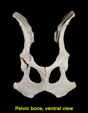
Ilium: The ilium represents the cranial half of the os coxae. It is divided into a flat cranial part, the wing and a narrow caudal part, the body. Along its cranial border is a roughened area known as the iliac crest. The tuber coxae contains the cranial ventral iliac spine and the adjacent part of the ventral border of the wing of the ilium. The tuber sacrale is composed of the cranial and caudal dorsal iliac spines and the intervening bone. Running along the ventromedial edge of the sacropelvic surface of the body of the ilium is the arcuate line between the auricular surface and the iliopubic eminence of the pubis.
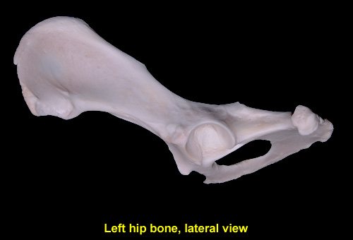
Ischium: The ischium consists of tuberosity, body, table, and ramus. It enters into the formation of the acetabulum, obturator foramen, and symphysis pelvis. The thick ischiatic tubrerosity provides attachment for the sacrotubeorus ligament. The ischiatic spine is a rounded crest dorsal to the acetabulum, where the ischium meets the ilium. The ramus of the ischium is the thin and wide fused with the pubis cranially. The left and right rami meet at the pelvic symphysis. The caudal border of each ischium meets to form the ischiatic arch.
Pubis: The pubis extends medially from the ilium and ischium to the symphysis. It consists of a body and two rami. On the cranial border of the cranial ramus of each pubic bone projects the iliopubic eminence. The cranial area of the pubis between the iliopubic eminences represents the pectens. From the midline of the pubis project the pubic tubercle.
The acetabulum has a semilunar articular surface that receives the head of the femur. Its circumference is broken at the acetabular notch. The ligament of the head of the femur attaches to the acetabular fossa.
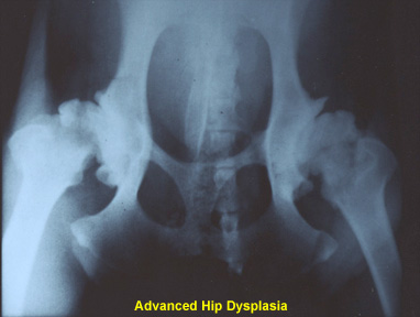
Clinical Correlation: Fractures of the pelvis are relatively common, and in many veterinary practices they constitute 20 to 30 percent of all fractures. Most fractures are multiple and involve more than one bone. Hip dysplasia is a hereditary disease that cause lameness and arthritis of the joints. It is caused by several contributing genetic factors, including abnormal articulation of the hip and femur causing friction, wear and tear within the joint. Hip dysplasia affects large and giant breeds of dogs.